CerebroVis: designing an abstract yet spatially contextualized cerebral arteries network visualization
Abstract
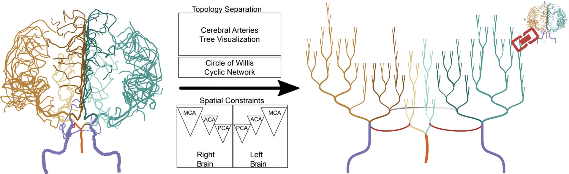
Blood circulation in the human brain is supplied through a network of cerebral arteries. If a clinician suspects a patient has a stroke or other cerebrovascular condition, they order imaging tests. Neuroradiologists visually search the resulting scans for abnormalities. Their visual search tasks correspond to the abstract network analysis tasks of browsing and path following. To assist neuroradiologists in identifying cerebral artery abnormalities, we designed CerebroVis, a novel abstract-yet spatially contextualized-cerebral artery network visualization. In this design study, we contribute a novel framing and definition of the cerebral artery system in terms of network theory and characterize neuroradiologist domain goals as abstract visualization and network analysis tasks. Through an iterative, user-centered design process we developed an abstract network layout technique which incorporates cerebral artery spatial context. The abstract visualization enables increased domain task performance over 3D geometry representations, while including spatial context helps preserve the user's mental map of the underlying geometry. We provide open source implementations of our network layout technique and prototype cerebral artery visualization tool. We demonstrate the robustness of our technique by successfully laying out 61 open source brain scans. We evaluate the effectiveness of our layout through a mixed methods study with three neuroradiologists. In a formative controlled experiment our study participants used CerebroVis and a conventional 3D visualization to examine real cerebral artery imaging data to identify a simulated intracranial artery stenosis. Participants were more accurate at identifying stenoses using CerebroVis (absolute risk difference 13%). A free copy of this paper, the evaluation stimuli and data, and source code are available at https://osf.io/e5sxt.
Materials
PDF | Preprint | DOI | Homepage | Supplement | Code | Demo | Video Preview | Demo Video | Demo Video (Archive) | BibTeXAuthors

Harsh Shukla
Geoffrey S. Young
Lei Qin
Amir A. Zamani
Liangge Hsu
Raymond Huang


Citation
CerebroVis: designing an abstract yet spatially contextualized cerebral arteries network visualization
Aditeya Pandey, Harsh Shukla, Geoffrey S. Young, Lei Qin, Amir A. Zamani, Liangge Hsu, Raymond Huang, Cody Dunne, and Michelle A. Borkin. IEEE Transactions on Visualization and Computer Graphics—VIS/TVCG. 2019. DOI: 10.1109/TVCG.2019.2934402
PDF | Preprint | DOI | Homepage | Supplement | Code | Demo | Video Preview | Demo Video | Demo Video (Archive) | BibTeX
Khoury Vis Lab — Northeastern University
* West Village H, Room 302, 440 Huntington Ave, Boston, MA 02115, USA
* 100 Fore Street, Portland, ME 04101, USA
* Carnegie Hall, 201, 5000 MacArthur Blvd, Oakland, CA 94613, USA